Pisum Genetics
2010-Volume 42
Research Papers
Contributions to the characterization of Vavilovia formosa (syn. Pisum formosum.). I. Anatomy of stem, leaf and calyx
Zoric, L. 1, Lukovic, J. 1, University of Novi Sad, Faculty of Sciences, Novi Sad, Serbia
Mikic, A. 2, Akopian, J.3, 2Institute of Field and Vegetable Crops, Novi Sad, Serbia
Gabrielyan, I. 3, 3National Academy of Sciences, Institute of Botany, Yerevan, Armenia
Sarukhanyan, N.4, Vanyan, A. 4, 4Green Lane Agricultural Assistance NGO, Yerevan, Armenia
Smykal, P.5, Kenicer, G.6 , 5Agritec Plant Research Ltd., Sumperk, Czech Republic
Vishnyakova, M.7 and 6Royal Botanical Garden Edinburgh, Edinburgh, UK
Ambrose, M.8 7N. I. Vavilov Institute of Plant Industry, St. Petersburg, Russia
8John Innes Centre, Norwich, UK
It was recently reported that Vaviloviaformosa Stev. Fed., informally referred to as beautiful vavilovia or simply vavilovia, has been gaining interest since its importance for legume molecular taxonomy and potential use in breeding as a useful crop wild relative (1). This is not surprising since it is a relict and endangered species limited to the Caucasus and its adjacent regions (2). As the only species of the genus Vavilovia Fed., it is often considered the closest to the common ancestor of the tribe Fabeae (syn. Vicieae) (3), comprising vetchlings (Lathyrus L.), lentils (Lens Mill.), peas (Pisum L.) and vetches (Vicia L.).
V. formosa is a perennial herbaceous species, with a dwarf habit and plant height between 5 and 15cm. Its stems are slender, sprawling or creeping, not winged and with a glabrous surface. The leaf of V. formosa is compound as in all other Fabeae with small, semi-sagittate and foliaceous stipules, one pair of broadly, cuneate-obovate to suborbicular, thick and glabrous leaflets with non-indented margins and the rachis ending with a mucro-like formation similar to faba bean (4).
Little is known about the anatomy of vegetative and reproductive organs of V. formosa. The aim of this preliminary research was to gain knowledge of the anatomical characteristics of V. formosa in order to properly understand the biology, ecology, adaptation strategy and nutritive value of this species.
Materials and Methods
On July 17, 2009, the first of the three expeditions was carried out to the Mount Ughtasar region in southern Armenia aimed at in situ research and ex situ conservation of V. formosa in Armenia. The Mount Ughtasar region is one of the three main habitats of V. formosa in Armenia (5). During the expedition, samples of stems, leaves and flowers of one population growing at an altitude of between 3305 and 3315 masl were collected. The collected fresh plant material was fixed in Carnoy I, a 3:1 solution of ethanol and glacial acetic acid.
The anatomical structures of stems and leaves, including the structure of leaflets, stipules, petiole and the rachis tip were observed using light microscopy and an Image Analyzing System, Motic 2000 (Motic, Wetzlar, Germany). Analysis of the calyx was added since fresh and properly fixed floral material was available. Cross sections of both aboveground vegetative organs and the calyx were made using a Leica CM 1850 cryostat (Leica, Solms, Germany) at a temperature of -18 to - 20 °C and at cutting intervals of 25 um.
Results
Stem. The stem is rounded in cross section, with two small wings, which are not prominent (Fig. 1). The primary cortex is well developed, with two layers of chlorenchyma cells and several layers of parenchyma cells without chloroplasts. No collenchyma is recorded. Two cortical vascular bundles, one in each wing,
21
Pisum Genetics
2010-Volume 42
Research Papers
also occur. In the central cylinder, collateral open vascular bundles of
Figure 1. V. formosa stem cross section
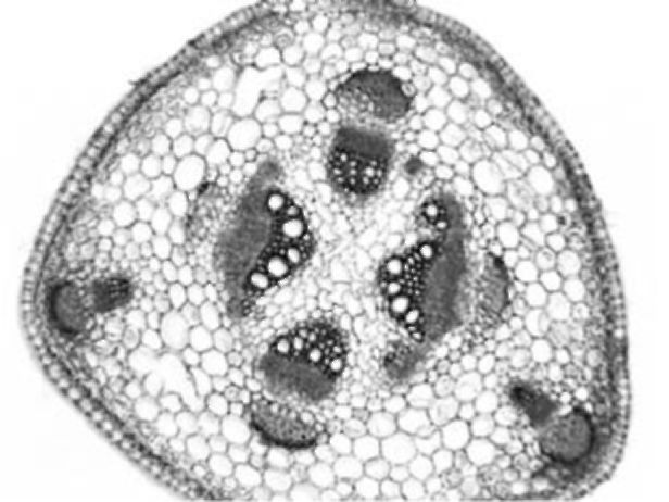
different size are arranged in a circle. Only small groups of sclerenchyma are present near the bundles. Sclerenchyma cells adjacent to the phloem have larger lumen and less lignified walls. Crystals occur in parenchyma sheath cells above the sclerenchyma strands. The central cylinder is composed of parenchyma with no central cavity or sclerenchymatic parenchyma present. Parenchyma cells of the cortex and central cylinder sometimes contain starch grains.
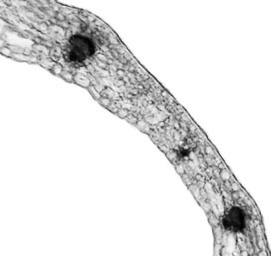
Leaflet. The leaflets of V. formosa have dorsiventral structure (Fig. 2). The
epidermis has a single layer composed of cells with sinuous anticlinal walls and covered with a thick cuticle. It contains large numbers of stomata on both the adaxial and abaxial surface. Stomata are slightly
sunken or at the same level as other epidermal cells. The leaves
Figure 2. V. formosa leaflet cross section
are glabrous on the adaxial side, but rare non-glandular trichomes were observed abaxially along the leaf veins. Leaf photosynthetic tissue is differentiated on palisade and spongy tissue. Palisade tissue is well developed, composed of two to three layers of elongated cells. Under the abaxial epidermis 4-5 layers of spongy tissue cells of irregular shape were observed. Mesophyll cells were closely arranged with small intercellular spaces. Closed collateral vascular bundles were arranged in a single row. Parenchyma sheath cells along the vascular bundles had solitary prismatic crystals. They are larger by the vascular bundle in the main vein which is not very prominent on the abaxial side. Collenchyma tissue is not present or only a few collenchyma cells occur abaxially.
Stipule. Stipules have anatomical structure very similar to the structure of the leaves (Fig. 3). Palisade tissue is not as dominant as the one in the leaves. It is composed of one or two layers of cells. Spongy tissue is present in three to four layers. Larger groups of sclerenchyma tissue are recorded by vascular bundles with solitary crystals in the sheath cells. Stipules are also glabrous.
Figure 3. V. formosa stipule cross section
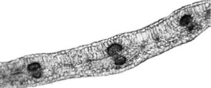
Figure 4. V. formosa petiole cross section
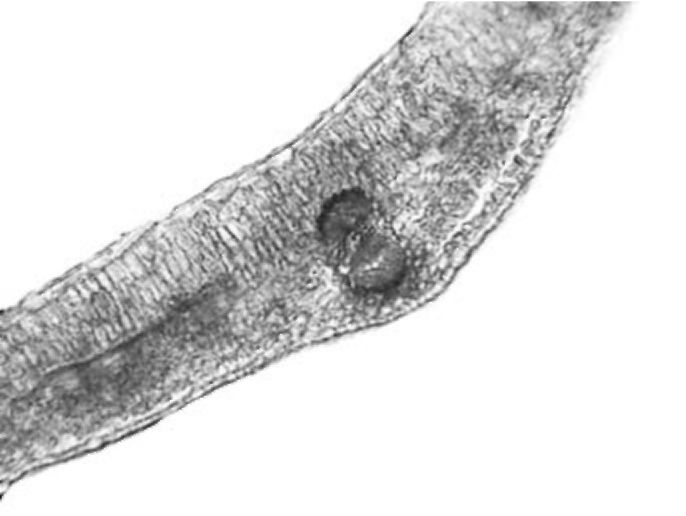
Petiole. The petiole is triangular or heart-shaped in cross section (Fig. 4). The epidermis has a single layer with sparse non-glandular trichomes. Four to five layers of chlorenchyma tissue occur subepidermally. Collenchyma is present in small groups only in the main rib. Five collateral vascular bundles are arranged in the form of an open arc. Sometimes, three vascular bundles in the main ribs are much larger than the other two. Small groups of sclerenchyma tissue occur by the phloem and are somewhat larger by the xylem. Solitary crystals were observed in parenchyma sheath cells above the vascular bundles. The central part of the petiole is filled with parenchyma cells. The central cavity is not present.
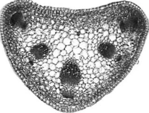
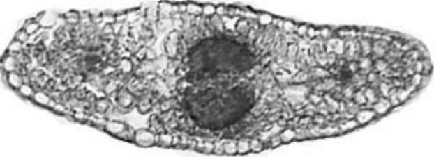
Figure 5. V. formosa rachis tip cross section
Rachis tip. The cross section of a V. formosa rachis tip is elliptical in shape (Fig. 5). On the adaxial side one layer of palisade cells is present. Mesophyll is compact with densely distributed cells.
22
Pisum Genetics
2010-Volume 42
Research Papers
Three vascular bundles, one large in the middle and one smaller on each side, are present in the mesophyll. Sclerenchyma tissue is recorded by the phloem and xylem and crystals are visible in vascular bundle sheath cells.
Figure 6. V. formosa calyx cross section
Calyx. The calyx has an egg-like shape in cross section (Fig. 6).
A thick cuticle covers the outer epidermis. Non-glandular and glandular trichomes are present on the inner epidermis. Non-glandular trichomes are thin, long, composed of one basal and elongated terminal cell. Three types of glandular trichomes are detected: Type I with one stalk cell and elongated multicellular secretory head, Type II with one stalk cell and a two-celled secretory head and Type III with a two-celled stalk and two-celled secretory head. Mesophyll was composed of 3-5 (or 7) layers of cells. Ten larger vascular bundles and several smaller between them are arranged in a circle. Crystals are present in
parenchyma cells adjacent to the vascular bundles.
Discussion
The results of anatomical analysis showed that V. formosa has general anatomical structure that is characteristic for most of the plants of family Fabaceae (6). However, this species developed some anatomical modifications, according to the environmental conditions of the specific habitat where it grows. It inhabits high mountain areas and grows on slopes with sparse vegetation coverage (5, 7). The slopes with V. formosa populations are sun-warmed and exposed to sunlight. The plant organ that reacts most to the environmental factors, especially the amount of sunlight, is the leaf (8). The leaves of V. formosa had helio-xeromorphic structural adaptations. They were relatively thick, covered with a thick cuticle, with slightly sunken stomata and with strongly developed palisade tissue. Furthermore, palisade tissue cells were elongated, narrow and arranged in three rows, which were adaptations that enabled this species to survive in this specific habitat.
According to the previous morphological descriptions of the V. formosa stem, it is slender, sprawling or creeping and without wings (5, 9). However, anatomical investigations of stem structure showed that stems had lateral wings which were not prominent but contained one cortical vascular bundle each. These wings were hard to observe by morphological examination only; they looked more like small stem ribs, but on cross sections the presence of wing bundles was obvious. The presence of a creeping stem, assigned as thread-like rhizoma by some authors (7), could be explained by the fact that stem mechanical tissue was not well developed. Collenchyma tissue was not recorded and sclerenchyma cells formed small groups by the vascular bundles. Sclerenchyma cells adjacent to phloem had larger lumen and less lignified cell walls. This amount and pattern of distribution of mechanical tissue could not provide enough mechanical support to the stem to keep it in an upright position (10).
It was noticed that V. formosa had been frequently grazed by wild and domestic animals, which was one of the main problems for its conservation (5, 7). One of the reasons for intensive grazing may lie in the fact that all analyzed vegetative plant parts are practically glabrous, contain small amounts of indigestible mechanical tissue and large amounts of well developed parenchyma tissue, which are favorable forage characteristics for animal consumption. These anatomical features of V. formosa should be further analyzed as potential benefits that could be introduced to other cultivated Fabeae plants and close relatives of this wild-growing species.
23
Pisum Genetics
2010-Volume 42
Research Papers
Acknowledgements
This work was financially supported by the Ministry of Science and Technological Development, Republic of Serbia, Grants No. and 20090 and 31024.
The authors are grateful to Darko Znaor, Zvonimir Bede and Dusan Dozet for their assistance. References
1. Smykal, P., Kenicer, G.J. and Mikic, A. 2009. Book of Abstracts of the IV Congress of the Serbian Genetic Society, Tara, Serbia, 1-5 June 2009, p. 166.
2. Kenicer, G.J., Smykal, P., Visnhakova, M. and Mikic, A. 2009. Grain Legumes 51: 8.
3. Golubev, A.A. 1990. Bulletin for Applied Botany, Genetics and Plant Breeding 135: 67-75.
4. Mikic, A., Smykal, P., Kenicer, G., Vishnyakova, M., Akopian, J., Sarukhanyan, N., Gabrielyan, I., Vanyan, A., Toker, C., Cupina, B., Ambrose, M., Mihailovic, V. and Ellis, N. 2009. Pisum Genetics 41:
34-39.
5. Akopian, J., Sarukhanyan, N., Gabrielyan, I., Vanyan, A., Mikic, A., Smykal, P., Kenicer, G., Vishnyakova, M., Sinjushin, A., Demidenko, N. and Ambrose, M. 2010. Genetic Resources and Crop Evolution 57: 1127-1134.
6. Metcalfe, C.R. and Chalk, L. 1957. Anatomy of the Dicotyledons, Vol I. Clarendon Press, Oxford.
7. Akopian, J.A. and Gabrielyan, I.G. 2008. Crop Wild Relative 6: 26-27.
8. Dickison, W. C. 2000. Integrative Plant Anatomy. Academic Press, San Diego.
9. Mikic, A., Smykal, P., Kenicer, G., Sarukhanyan, N., Akopian, J., Gabrielyan, I., Vanyan, A., Sinjushin, A., Demidenko, N., Cupina, B., Mihailovic, V., Vishnyakova, M. and Ambrose, M. 2010. Field and Vegetable Crops Research 47: 387-394.
10. Petrova, M. V. 1973. Bulletin for Applied Botany, Genetics and Plant Breeding 51: 126-134.
24
Hosted by uCoz