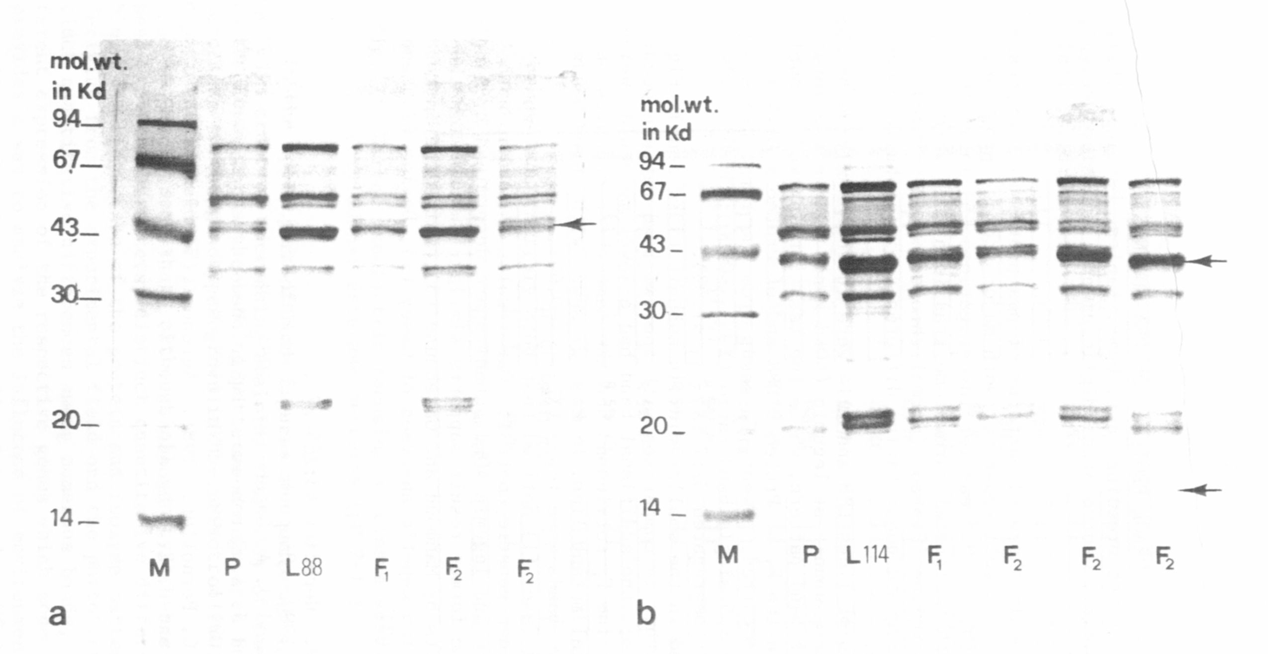
Fig. 1. SDS electrophoregrams of reduced globulins in parental lines, F1 and F2
M=molecular weight markers.
a) Proteo (P) and L88
b) Proteo (P) and L114
|
|
|||
|
PNL Volume 17 1985
|
RESEARCH REPORTS 63
|
||
|
|
|||
|
GENETIC ANALYSIS OF SEED STORAGE PROTEINS OF PEA MUTANTS1/
Rao, R., M.R. Mogno, Plant Breeding Lnst. Univ. Naples, Portici, Italy
and S. Grillo C.N.R., Centro Studi Miglioramento, Genetico Ortaggi,
Portici, Italy
Genetic variation in the band patterns of pea proteins as expressed
in gel electrophoresis is well known for both major storage proteins,
vicilin and legumin (6,7). Our earlier investigations (5) in which
eight Pisum lines with reciprocal chromosome translocations were
compared with the test line 'Proteo' (normal chromosome structure)
showed both qualitative and quantitative differences in globulin elec-
trophoretic profiles.
Storage proteins of lines L88 and L114 were further analyzed by SDS
electrophoresis. Theasubunit of legumin (40Kd zone) in the two mutant
lines showed a single band pattern, while the test line showed a double
band (Fig. la,b). In the F1 of Proteo x L88 and of Proteo x L114 a
double band pattern occurred. Twenty F2 seeds from the first cross seg-
regated in 15 double : 5 single band ratio (X23:1=0.00); 39 F2 seeds
from the second cross segregated 32:7 (X23:1) =1.03).
Line L114 showed in the smaller vicilin subunlt (15Kd zone) (Fig.
lb) three bands together in comparison with the two band pattern occur-
ring in the test line; the additional band had a lower molecular weight
than the other two. The F1 cotyledons had the same three subunits ob-
served in the parental mutant line. In F27 27 seeds with three bands
and 12 seeds with two bands were found (X23:1 =0.69).
Thomson (7) and Casey (1) noticed that both vicilin and legumin
band patterns are under genetic control. Our results show that both
Vicilin 15Kd subunits and legumin 40Kd subunits are determined by single
Mendelian genes. The former result supports similar findings for the
major Vicilin subunits by Mahmoud and Gatehouse (3), while the latter
result corroborates the genetic analyses of Casey (1), Davies (2), and
Matta and Gatehouse (3).
1. Casey, R. 1979. Heredity 43:265-272.
2. Davies, D. R. 1980. Biochem. Genet. 18:1207-1219).
3. Mahmoud, S. H. and J. A. Gatehouse. L984. Heredity 53:185-191.
4. Matta, N. K. and J. A. Gatehouse. 1982. Heredity 48:383-392.
5. Rao, R. and S. Grillo. 1984. PNL 16:68-69.
6. Rao, R. and J. C. Pernollet. 1981. Agronomic 1:909-916.
7. Thomson, J. A. and H. E. Schroeder. 1978. Aust. I. Plant Physiol.
5:281-294.
Ś^ Contribution no. 12 from Centro di Studio per il Migl ioramento
Genetico degli Ortaggi - C.N.R. - Portici (Napoli) - Italy
|
|||
|
|
|||
|
|
||
|
|
||
|
|
||
|
Fig. 1. SDS electrophoregrams of reduced globulins in parental lines, F1 and F2
M=molecular weight markers.
a) Proteo (P) and L88
b) Proteo (P) and L114
|
||
|
|
||